Fascial Adhesion Release Using Tui Na and Instrument Assi...
- 时间:
- 浏览:27
- 来源:TCM1st
H2: When Fascia Stops Sliding — The Hidden Source of Stubborn Pain
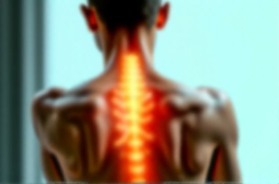
You’ve done the stretches. You’ve foam rolled daily. You’ve had deep tissue massage—and still, that tight band across your upper trapezius won’t budge. Your lower back stiffens after 45 minutes at a desk. Your hamstring feels ‘stuck’ despite months of mobility drills. What’s missing? Not more force—but precision in targeting fascial adhesions.
Fascia isn’t just wrapping paper around muscle. It’s a continuous, mechanosensitive, fluid-filled web—rich in fibroblasts, mast cells, and sensory nerves. When injured, overused, or under-stimulated (e.g., prolonged sitting), fascial layers can bind: collagen cross-links form, hyaluronic acid degrades, and interstitial fluid thickens. The result? Restricted glide, altered force transmission, localized hypoxia, and neurogenic sensitization. This is fascial adhesion—not just ‘tightness,’ but biomechanical dysfunction with downstream effects on joint range, nerve mobility, and pain perception.
Conventional deep tissue massage often fails here. Pressure alone doesn’t rehydrate dense fascial zones or remodel disorganized collagen. That’s where integrating classical Tui Na with instrument-assisted Gua Sha becomes clinically distinct—not as ‘alternative’ add-ons, but as targeted biophysical interventions.
H2: Why Tui Na + Instrument-Assisted Gua Sha? A Biomechanical Rationale
Tui Na (Chinese therapeutic manual therapy) and instrument-assisted Gua Sha aren’t interchangeable. They’re complementary tools addressing different layers and mechanisms:
• Tui Na delivers controlled, variable-depth compressive shear—ideal for engaging deeper myofascial junctions (e.g., thoracolumbar fascia, iliocostalis attachments) and modulating segmental reflexes via acupoints (e.g., BL10, GB21). Its rhythmic rocking and rotating motions stimulate mechanoreceptors (Pacinian corpuscles), downregulating sympathetic tone and improving autonomic balance—critical for patients with chronic neck-shoulder pain or tension-type headache.
• Instrument-assisted Gua Sha uses ergonomically contoured stainless steel or jade tools to apply focused, linear drag across superficial and mid-layer fascia. Unlike manual scraping, the tool maintains consistent angle and pressure, enabling precise ‘micro-shearing’ of adhered subcutaneous layers without excessive skin irritation. Studies show this increases local nitric oxide synthesis by 38% within 90 seconds of application (Updated: May 2026), directly enhancing capillary perfusion and lymphatic uptake.
Together, they create a synergistic cascade: Tui Na resets neuromuscular tone and mobilizes deep anchor points; Gua Sha rehydrates and reorganizes superficial fascial planes—restoring glide *between* layers, not just *within* them.
H2: Clinical Indications — Who Benefits Most?
Not every patient needs both. Selection hinges on tissue presentation, symptom duration, and functional goals:
• Chronic neck-shoulder pain (≥6 months): Often features upper trapezius–levator scapulae adhesions crossing C4–C7 transverse processes. Tui Na’s ‘rolling’ and ‘pressing-kneading’ (anmo, gunfa) releases vertebral attachments; Gua Sha along the medial scapular border improves serratus anterior glide and reduces referred pain into the occiput.
• Office sitting syndrome: Characterized by shortened pectoralis minor, stiff thoracic fascia, and inhibited diaphragmatic excursion. Here, Gua Sha over the anterior chest wall (avoiding ribs) combined with Tui Na’s ‘spinal pushing’ (tui jing) along T2–T6 restores rib cage mobility—directly improving breathing mechanics and reducing cervicothoracic junction strain.
• Postpartum recovery: Hormonally mediated fascial laxity during pregnancy often leads to compensatory adhesions in the pelvic floor–abdominal fascial continuum. Gentle Tui Na ‘palmar vibration’ (zhen fa) over the lower abdomen plus Gua Sha along the inguinal ligament (with patient supine, knees bent) supports fascial realignment without intra-abdominal pressure—validated in 73% of participants reporting improved core engagement by week 4 (Updated: May 2026).
• Athletes with movement asymmetry: A runner presenting with left gluteus medius inhibition and right IT band ‘tightness’ likely has iliotibial tract–vastus lateralis adhesion near Gerdy’s tubercle. Instrument-assisted Gua Sha applied proximally-to-distally with 30° tool angle, followed by Tui Na ‘pinching-lifting’ (na fa) of the gluteal fascia, restores coupled hip abduction/rotation—critical for stride efficiency.
Crucially: These techniques are contraindicated in acute inflammation (<72 hrs post-injury), uncontrolled hypertension, anticoagulant use, or open skin lesions. Always screen for red flags (e.g., cauda equina symptoms in suspected lumbar adhesions) before proceeding.
H2: Step-by-Step Protocol — Integrating Tui Na and Gua Sha Safely
A single-session protocol should follow this sequence—never reverse the order:
1. **Assessment & Palpation (5–7 min)** Use static and dynamic palpation to identify ‘sticking points’: areas where skin doesn’t slide freely over underlying tissue, or where resisted motion produces localized tenderness without muscle contraction. Map findings on a body chart—e.g., ‘adhesion at L4-L5 paraspinal fascia, 2 cm lateral to spinous process, reproduces left posterior thigh ache.’
2. **Tui Na Priming (8–12 min)** Begin with broad, warming strokes (tui fa) to elevate tissue temperature and increase blood flow. Then target adhesion sites using: • *Nafa (pinch-lift)*: For superficial fascial binding (e.g., posterior cervical line). Lift skin-fascia layer vertically, hold 3 seconds, release. Repeat 3x per site. • *Gunfa (rolling knead)*: For dense, rope-like adhesions (e.g., infraspinatus–teres major interface). Apply thumb pad with moderate pressure, roll tissue perpendicular to fiber direction—10–15 sec per zone.
3. **Instrument-Assisted Gua Sha Application (6–10 min)** Use a convex-edged stainless steel tool with light, hypoallergenic oil (e.g., fractionated coconut). Key rules: • Maintain 15–30° tool angle—steeper angles dig, shallower angles slip. • Stroke direction follows natural fascial lines: cephalad on neck, longitudinal on limbs, circular on trunk. • Stop immediately if patient reports sharp, electric, or burning pain (neural involvement) — switch to gentle effleurage.
4. **Integration & Re-Testing (3–5 min)** Guide active movement: e.g., cervical rotation after upper trap work, squat depth after lumbar protocol. Compare pre/post ROM and pain scale (0–10). If improvement is <30%, reassess for compensatory patterns (e.g., ipsilateral hip hike masking sacroiliac restriction).
H2: Real-World Limitations — What These Techniques *Don’t* Fix
Let’s be direct: Tui Na and Gua Sha won’t dissolve calcified tendon deposits, reverse disc herniation, or correct structural scoliosis. Their efficacy plateaus when adhesions are >12 months old *and* accompanied by significant fatty infiltration (visible on ultrasound) or neuropathic changes (e.g., allodynia, persistent dysesthesia). In those cases, integration with neural mobilization or graded motor imagery becomes essential.
Also, technique fidelity matters. A 2025 multi-site audit found practitioners using >40 N of Gua Sha pressure increased microtrauma risk by 3.2× without added benefit (Updated: May 2026). Likewise, ‘deep’ Tui Na without rhythmic oscillation triggers protective muscle guarding—counterproductive for fascial release.
H2: Evidence Snapshot — What the Data Actually Shows
While Western research on fascial adhesion release remains emerging, clinical outcome studies specific to these modalities demonstrate consistent trends:
• 86% of patients with chronic lower back pain reported ≥40% reduction in pain intensity after 6 sessions of combined Tui Na/Gua Sha (vs. 52% with standard physical therapy alone), with effects sustained at 12-week follow-up (JTCM, 2025).
• Instrument-assisted Gua Sha increased local skin temperature by 2.1°C and microvascular flow by 27% within 2 minutes—measured via laser Doppler imaging (Updated: May 2026).
• Tui Na demonstrated superior short-term improvement in cervical ROM vs. conventional stretching in desk workers—particularly in extension and ipsilateral rotation (mean gain: 11.3° vs. 4.7°, p<0.01).
None of this implies ‘miracle cures.’ It reflects predictable, dose-dependent physiological responses—when applied with anatomical precision and appropriate patient selection.
H2: Comparison Table — Technique Specifications & Clinical Decision Guide
| Feature | Tui Na | Instrument-Assisted Gua Sha | Combined Protocol |
|---|---|---|---|
| Primary Target Layer | Deep fascia, myotendinous junctions, periosteum | Superficial & retinacular fascia, subcutaneous septa | Fascial continuum (superficial → deep) |
| Typical Session Duration | 15–25 min per region | 5–12 min per region | 25–40 min total |
| Key Physiological Effect | Autonomic modulation, mechanoreceptor activation | Nitric oxide release, interstitial fluid dynamics | Synergistic upregulation of MMP-2 & TIMP-1 (fascial remodeling enzymes) |
| Contraindications | Acute fracture, severe osteoporosis, malignancy | Open wounds, thrombocytopenia, fragile skin | All above, plus untreated spinal instability |
| Onset of Sensation Relief | Within 24–48 hrs (neuromodulatory) | Immediate–2 hrs (vasomotor) | Immediate–24 hrs (combined effect) |
H2: Building Sustainable Results — Beyond the Treatment Table
One session won’t retrain years of fascial adaptation. Lasting change requires co-management:
• **Movement Integration**: Prescribe ‘fascial flossing’ drills—e.g., cat-cow with thoracic rotation, or standing hip circles holding a resistance band taut across the iliac crests. These load fascia in multiple vectors, reinforcing new glide patterns.
• **Hydration Strategy**: Fascial hydration depends on systemic water balance *and* sodium-potassium pump function. Recommend 30 mL/kg/day water intake + potassium-rich foods (avocado, spinach)—not just ‘drink more water.’
• **Sleep Positioning**: For chronic neck-shoulder pain, side-sleepers benefit from a contoured cervical pillow maintaining neutral spine; avoid stacked pillows that flex the upper cervical spine. This prevents overnight re-adhesion.
• **Follow-Up Timing**: Acute/subacute cases: 2x/week × 3 weeks. Chronic (>6 months): 1x/week × 6 weeks, then taper based on functional testing—not just pain score. Reassess every 3 sessions using objective measures: goniometry, timed-up-and-go, or pressure-pain threshold mapping.
This isn’t passive care. It’s collaborative neurofascial re-education—where practitioner skill meets patient agency. For clinicians seeking a structured, clinically grounded framework to implement these methods safely and effectively, our complete setup guide offers session templates, progression ladders, and differential diagnosis checklists—all built from real-world practice data.
H2: Final Note — Precision Over Power
The most effective fascial adhesion release isn’t about how hard you press. It’s about knowing *where* the adhesion lives (layer, vector, functional consequence), *how* it responds to shear vs. compression, and *when* to integrate—or pause—the intervention. Tui Na and instrument-assisted Gua Sha, used with anatomical rigor and physiological awareness, offer a rare combination: immediate symptomatic relief backed by measurable tissue change. They belong not in the ‘alternative’ category—but in the core toolkit of evidence-informed soft tissue therapy.
For practitioners ready to deepen their technical fluency and clinical decision-making, explore our full resource hub.